The Coutinho-Budd lab is interested in the cellular and molecular aspects of glial cell development and function. Glia are critical components of the nervous system—almost everything that neurons do requires glial support. Significant progress has been made in understanding neuron-glia interactions at synapses and axons, but how glia support neuronal cell bodies remains poorly defined.
Many glial cell types in the mammalian nervous system have subsets that interact with neuronal cell bodies (e.g. protoplasmic astrocytes, satellite microglia, and perineuronal oligodendrocytes in the brain and spinal cord, Müller glia in the retina, and satellite glia in peripheral ganglia). Unfortunately, the genetic tools needed to study glial interactions only at neuronal cell bodies don’t exist in mammals.
My lab takes advantage of Drosophila cortex glia—a remarkable subclass of glia that extend fine processes to individually wrap almost every neuronal cell body in the central nervous system—as a model to study these neuron-glia interactions in vivo. Cortex glia are thought to secrete factors for neuronal development, buffer ions and nutrients in the extracellular environment, and remove debris during development and after injury. Animals with dysfunctional cortex glia exhibit behavioral deficits ranging from poor coordination and impaired locomotion to seizures, and a lack of cortex glia can be lethal, yet these cells have not been extensively studied. My lab aims to better understand how cortex glia develop, communicate with surrounding neurons and glia, and contribute to overall nervous system function.
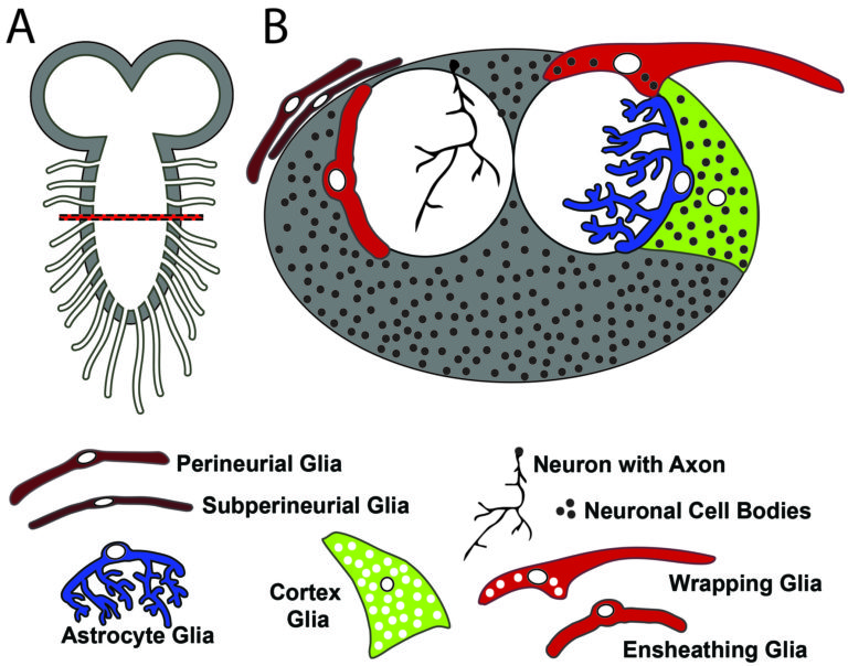
A) The Drosophila (aka fruit fly) larval central nervous system (CNS) contains two brain lobes and a ventral nerve cord (akin to the spinal cord), with nerve bundles exiting towards the body. B) Cross section of the ventral nerve cord along the horizontal line in (A). The Drosophila CNS is divided into two main areas: the cortex (in gray) that contains neuronal cell bodies, and the neuropil (in white) that contains the axons and synapses. One example neuron is shown with its cell body in the cortex, and it's neurites extending to the neuropil. The Drosophila CNS also has many glial subtypes: the surface glia that make up the blood brain barrier (Perineurial and subperineurial glia), cortex glia that predominantly wrap the neuronal cell bodies, ensheathing glia that encase the neuropil, astrocytes that extend cellular processes into the synaptic neuropil, and wrapping glia that predominantly wrap axon bundles.
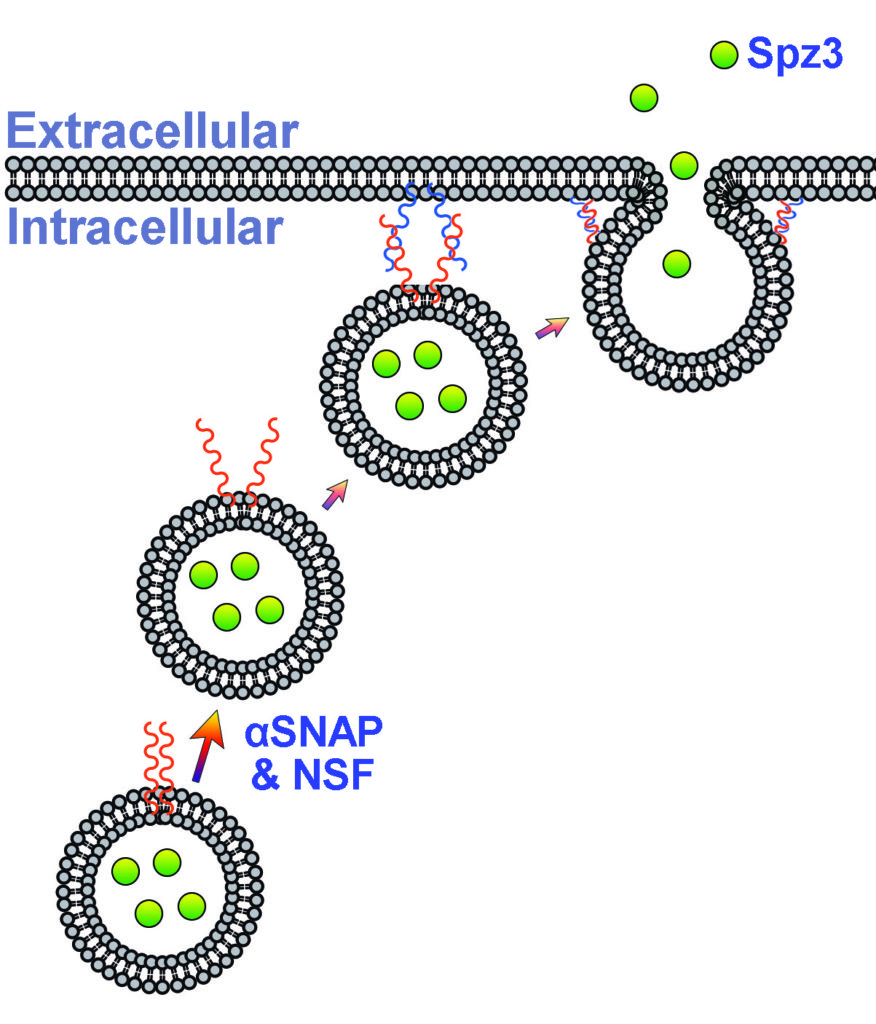
A) Diagram of vesicle during exocytosis. In order for a vesicle to fuse to a target membrane, cis-SNARE proteins (orange) must unzip via αSNAP and NSF to bind to the target SNARE proteins (blue), allowing the vesicle to fuse with the target membrane and release its contents, such as a the secreted protein Spz3 (green).
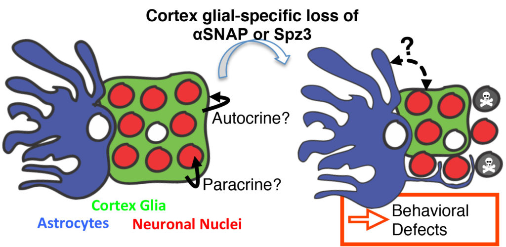
Normal cortex glia wrap neuronal cell bodies in the cortex, forming a tight border and boundary with astrocytes at the neuropil. After the loss of either αSNAP or Spz3 from cortex glia, the cortex glia lose their ability to wrap neurons, and instead form globular structures leaving many neurons unwrapped. This leads to increased neuronal cell death, aberrant invasion of astrocyte processes from the neuropil into the cortex, and ultimately behavioral impairment such as poor locomotion. Future projects aim to better understand exactly how this impaired wrapping causes such wide ranging effects, and what are the molecular cues that underly the glial-glial communication and neuron-glia crosstalk.
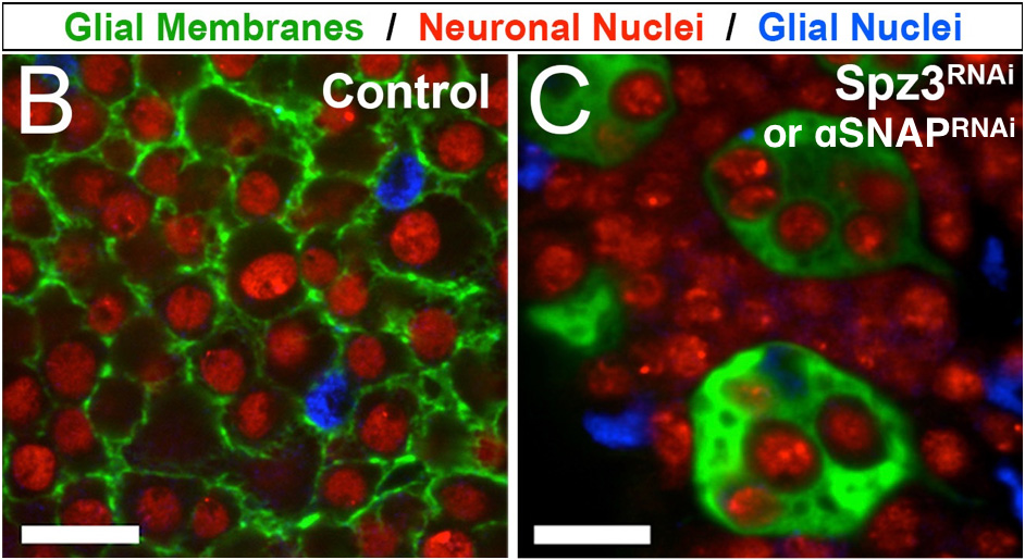
Left: Close-up of thin cortex glial processes (green) wrapping individual neuronal cell bodies (neuronal nuclei in red), with glial nuclei in blue. Right: Loss of either αSNAP or Spz3 results in globular cortex glia, leaving many neurons unwrapped, and some neurons encased within the cortex glial spheres.
One approach in understanding how neurons and glia communicate is to use biosensors to visualize molecules in real time in vivo (i.e. living brains). Here, a calcium indicator expressed in cortex glia (green) shows calcium signaling events occurring in the fine processes that surround neuronal cell bodies (red).